RecommendMail Facebook LinkedIn
Explore your micro universe with JENOPTIK GRYPHAX® cameras for micro photography
Making the hidden visible: in Life Science & Medical Science, Material Science & Manufacturing, Education
The fascination with the unknown drives human curiosity. The strong drive to explore things motivates us to come up with new ideas. It encourages us to comprehend the intricacy of our world in the past and present and to positively influence it in the future. Making the hidden visible with the help of micro photography enables the constant renewal of individual building blocks of knowledge and the ongoing pursuit of completing a puzzle. This is how our overall picture is perpetually being updated.
Jenoptik brings together the curious people, their technical knowledge, their application know-how and their pioneering spirit. This combination of precision and intelligence creates products and solutions which set impressive standards.
JENOPTIK GRYPHAX® cameras provide those who are searching for answers to their questions with a solid tool to gain new insights and to examine tiny details.
Discover unknown galaxies of the micro universe
The passion to bring the hidden into the light is inherent in both the users of Jenoptik's microscope cameras and that fantastic being called GRYPHAX. With the joint mission to explore unknown galaxies of the micro universe, the GRYPHAX accompanies the curious people, the researchers, the teachers and the innovators on their daily journey of discovery.
GRYPHAX combines brilliant vision, a huge wingspan, dynamic maneuverability, bounce and surefootedness.

JENOPTIK Gryphax
Ease of use -
giving your work an effortless feel.
- User-friendly and intuitive microscope camera software
- Optimized workflows
- Live-image optimized, all features in real time, all the time
- Time-saving and easy installation
High Image Quality -
giving you the details to make the right decisions.
- Professional Jenoptik image quality, based on real measured colors by use of color-co-site sampling & microscanning
- Perfect color reproduction by use of spectrally measured sensors
Stability -
giving you a reliable research tool you can count on.
- USB 3.0 camera interface
- Secure investment: long-lasting & reliable hardware
- Secure investment: long-term software support & operating system compatibility
Versatility -
giving you the freedom to work with your favorite equipment.
- Cross-platform compatible for WIN, MAC & Linux
- Identical GUI across WIN, MAC & Linux platform
- Suitable for all microscope brands
- Free SDK available
- Twain, Direct X® and 3rd party software support
IMAGE QUALITY -
GRYPHAX' eagle eyes do not miss a single detail
Extraordinary vision as well as the ability to perfectly detect the tiniest color detail are the GRYPHAX®'s key features. Based on JENOPTIK's true-color know-how and spectrally measured sensors, JENOPTIK GRYPHAX® cameras deliver brilliant image quality in micro photography.
JENOPTIK GRYPHAX® cameras ensure brilliant low-noise image results, which provides a reliable basis for your decision-making. If your decisions depend on the tiniest details and color gradients, then the value of your work will be ultimately defined by a good camera. You should be able to see all the valuable image details provided by your microscope on your monitor screen with almost no image loss.
EASE OF USE -
GRYPHAX's powerful wings make your image analysis fly
The magic is in the details. This does not only apply to the smallest building blocks of knowledge which scientists constantly use to complete the overall picture. This also goes for the workflow that leads to the goal. GRYPHAX's large wings carry you effortlessly through your micro universe.
JENOPTIK GRYPHAX® cameras pave your way, the path that has to be followed in order to arrive at the individual results and the new findings. Sophisticated features of the JENOPTIK GRYPHAX® software included in the package contents such as auto brightfield setup, panorama or z-stacking tool facilitate image aquisition and analysis. User-friendly workflows will inspire your day-to-day activities.
VERSATILITY -
GRYPHAX's on-call agility ensures flexible manoeuvrability
Flexibility can be a great advantage in a dynamic environment. The muscular, cat-like GRYPHAX® body reflects its instantly adaptable agility and resilience. GRYPHAX's remarkable power provided by versatile software options allows flexible agility of your microscopy system.
JENOPTIK GRYPHAX® microscope cameras are the right choice when it comes to flexibility. The adaptability of JENOPTIK GRYPHAX® cameras gives you the freedom to work with your favorite equipment - no matter which brand of microscope, no matter which operating system. In addition to the GRYPHAX® software included in the delivery, there are further possibilities to integrate 3rd-party software, free drivers or a free SDK.
STABILITY -
GRYPHAX's strong paws give confident grip
A healthy dose of curiosity, patience and confidence is required to evaluate facts or validate a new hypothesis. The strong, serrated paws of the GRYPHAX® symbolize an unparalleled firm grip that provides you with the necessary stability to break new scientific ground.
JENOPTIK GRYPHAX® cameras offer you the required foothold for responsible decision-making. In addition to knowledge and attention to detail, it is the precision and reliability of the work tools that provide secure foothold. Highly reliable hardware and stable software as well as the finest reproducible image details and color gradations guarantee the stability which will help you get the job done with confidence.
Explore your micro universe


Application Report | Blog
How do doctors use optical technology to diagnose blood disorders? Hear it from an expert.
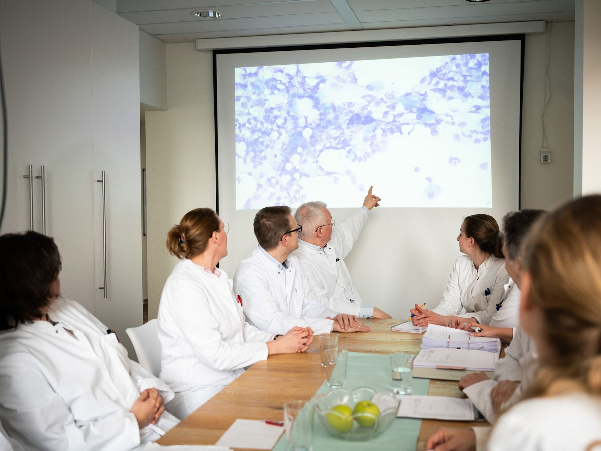
Precise hematological diagnosing requires a lot of experience and excellent true-color imaging from the microscope camera used for hematopathology – such as our GRYPHAX® ARKTUR camera.
We interviewed an expert, Thomas Nebe, M.D., from the HaeMa laboratory in Mannheim, Germany to get a better understanding of the benefits of precise imaging for hematopathology diagnoses.
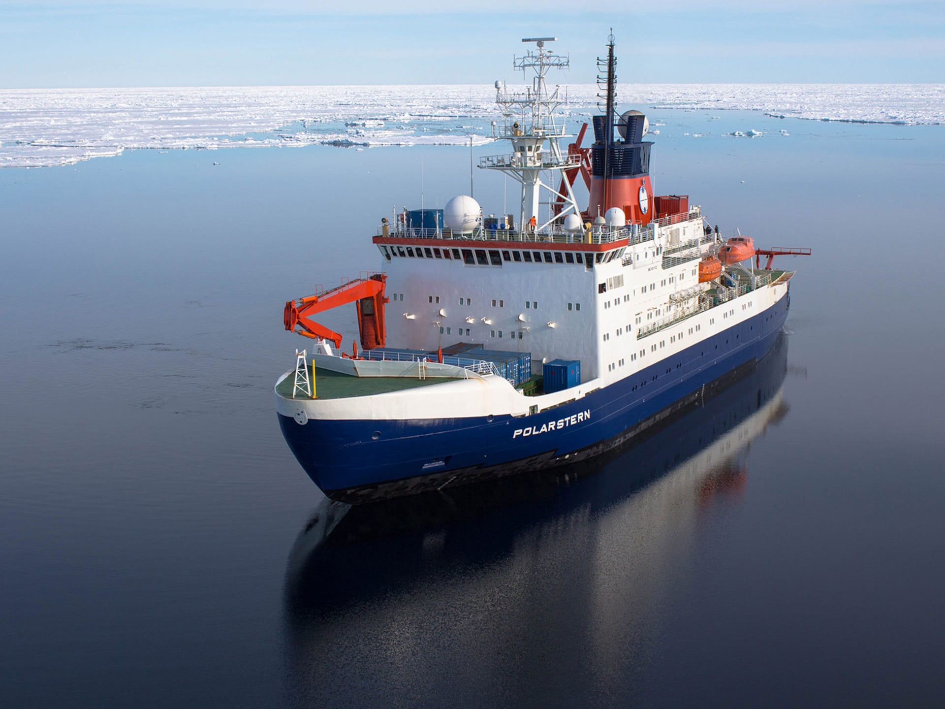
Blue depths and endless expanses – oceanography exposes the unknown

Marine researchers at the University of Hawaii use a JENOPTIK GRYPHAX® camera to document their discovery of a new type of deep-sea bamboo coral – insights into the application of an ALTAIR camera from Jenoptik for oceanography.
It is important to understand the phenomena of our oceans; they have far-reaching effects on our climate, our health and the economy. Our oceans are probably the least understood and yet one of the most important components of our planet.
The magic is in the details
Every discovery and decision that is made can change the world – sometimes only in tiny nuances or for a particular person – but sometimes for a great number of people, which could also have an enormous impact on our daily lives.
Read about an example of how a JENOPTIK GRYPHAX® camera is used to analyze tiny details in marine microfossils, to decode the data hidden in the sediments of our oceans and to reconstruct the environmental conditions in the course of the earth's climate history.
Genuine leather or imitation? In pursuit of quality with microscopy

Opinions and standards regarding the authenticity and naturalness of the material leather occasionally differ, particularly as the result of a globalized and increasingly online-driven trade.
This is where the identification of genuine leather can help to dispel any misunderstandings between manufacturers, retailers, purchasing companies and, not least, the consumers.
FIND OUT MOREMicroscope images help archaeologists to solve the mystery of the Griffin Warrior treasures

Researchers from the University of Cincinnati have conducted an archaeological analysis of a gold signet ring from the “Griffin Warrior Tomb” – one of the 3,500-year-old artifacts from excavations carried out at the Palace of Nestor in Pylos. Microscope images taken with JENOPTIK GRYPHAX® cameras form the basis for the digital investigations of the artefacts recovered from a Bronze Age time capsule.
The Palace of Nestor was described in Homer’s Odyssey and in the Greek myth of the Trojan horse and was an important administrative and political centre in the Mycenaean period. It is located in the ancient city of Pylos, on the southwest coast of Greece. In 2015, archaeologists from the University of Cincinnati made an exceptionally rich find at the Palace of Nestor. An initially inconspicuous rectangle made of stones revealed the marvelously intact tomb of a warrior from the Bronze Age.
Exploding the myths of digital microscopy: Is CCD always better than CMOS?
CMOS sensors are very attractive and have such an excellent price-performance ratio that virtually any device today can be equipped with the ability to image – and give any microscope, scientific instrument or medical device a significant competitive advantage. The ability to visualize samples – whether central to the analysis or an apparent “nice to have” feature – can lead to desirable new applications, quality assurance and experimental design.
Biological imaging has evolved from a passive observational collector of descriptive pictures to a keen, versatile and quantitative analytical tool. Microscopic images of tissues and cells provide the basis for characterization and measurements of disease progression, live cell imaging, automated diagnostics, and a host of other activities in the life science and medical diagnostic laboratory. Digital microscopes thus play a crucial role in the signal pathway between the biological sample of interest and the eyes of the scientist.
In Applications of Chemiluminescence, Can CMOS replace CCD? The advantage of replacing CCD with CMOS for applications where every photon is important.

CCD or CMOS? Although both devices are built on the same fundamental technology, there are differences that have favored one over the other, depending on the application. Chemiluminescence has often been the reserve of CCD detectors because of their relative sensitivity, but they are slower compared to CMOS. Now, CMOS has taken a great leap, and may be favorable to support the more challenging applications where readout speed, power consumption and portability are of the essence.
Chemiluminescence – the emission of light from a chemical reaction – is good for more than glow sticks and lecture demonstrations. In biological research, chemiluminescence informs scientists of the precise locations and quantities of proteins and metabolites within cells as well as in in vitro analysis methods such as Western blotting. Chemiluminescence provides key insights into the expression and activity of biologically significant proteins, which is often studied in the process of drug development.
Find out more about the microscope cameras:


EditContact